
Parameter
Mehr zum Buch
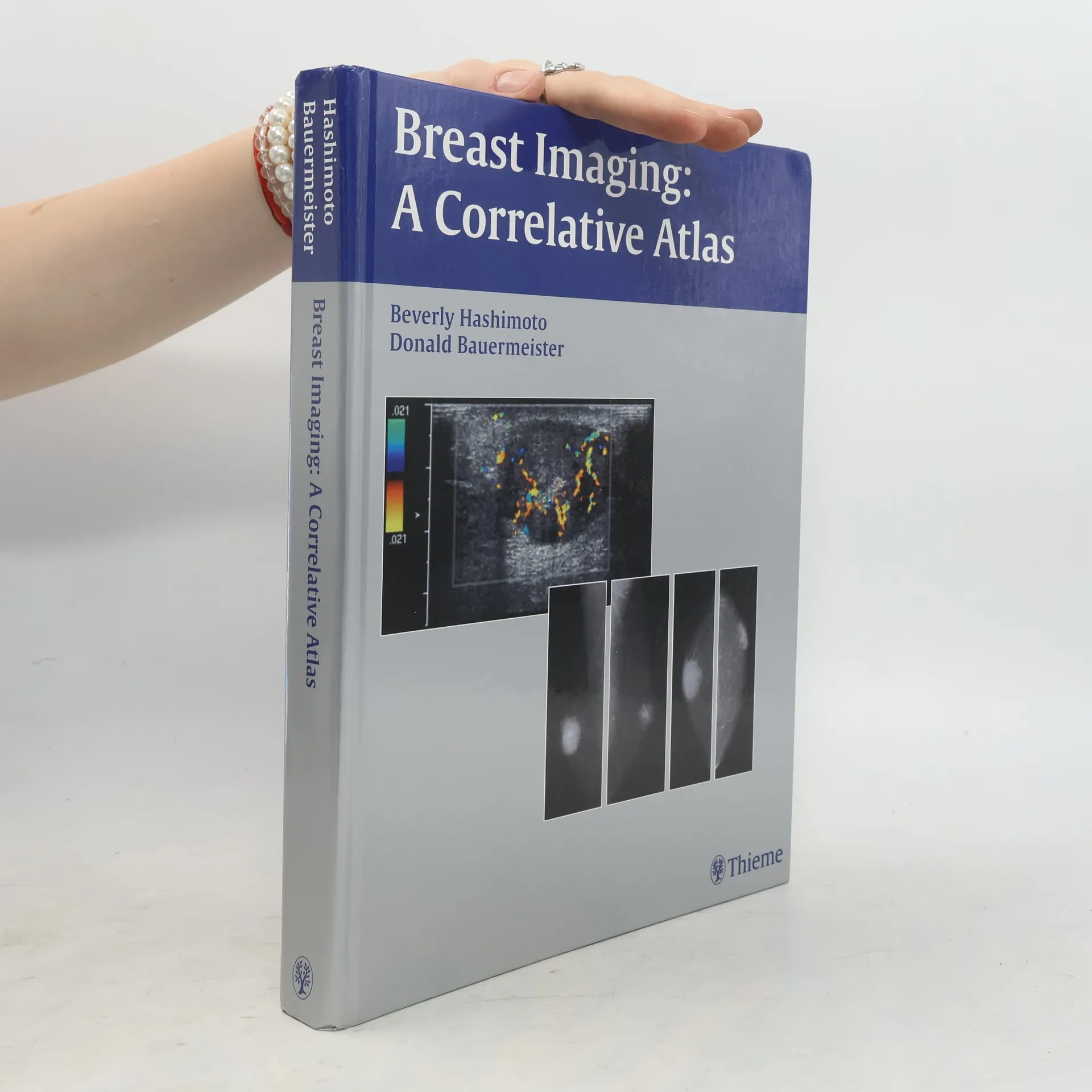
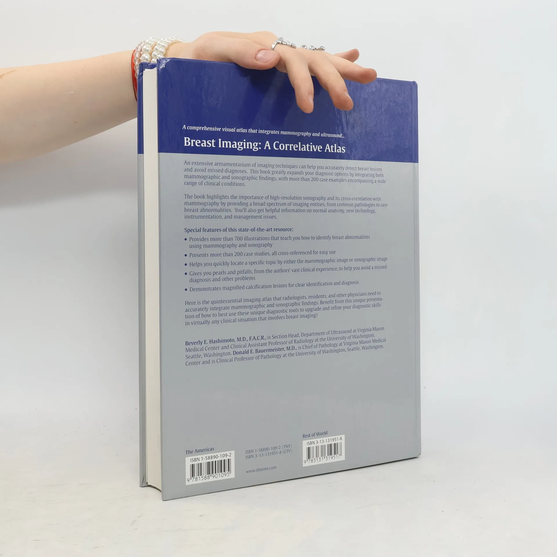
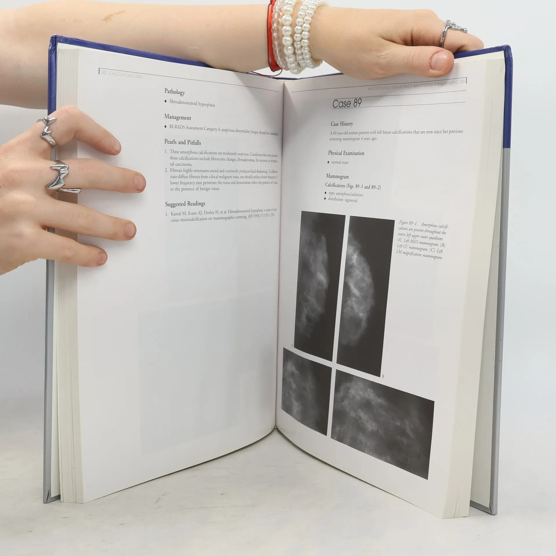
The book is targeted for breast imagers who wish to improve their interpretative skills by learning a pattern approach method to analyze and integrate mammographic and sonographic findings. One goal of the book is to emphasize the multidisciplinary nature of breast imaging utilizing sonography, magnetic resonance and various nuclear medicine techniques. Another goal is to demonstrate the importance of high-resolution sonography. The final objective of this book is to provide an atlas of a wide variety of pathological entities within the breast. It includes coverage of normal anatomy and variants, technology and instrumentation, histology for selected cases, and management. Two table of contents allow readers to access suspected diagnosis by either mmaography or ultrasound findings. The first enables readers to identify the histologic entities that create specific mammographic findings; the second aloows readers to study the lesions that produce sonographic abnormalities.
Buchkauf
Breast imaging, Beverly Hashimoto, Donald E. Bauermeister
- Sprache

- Erscheinungsdatum
- 2003
- product-detail.submit-box.info.binding
- (Hardcover)
Keiner hat bisher bewertet.
- Titel
- Breast imaging
- Untertitel
- A Correlative Atlas
- Sprache
- Englisch
- Autor*innen
- Beverly Hashimoto, Donald E. Bauermeister
- Verlag
- Thieme
- Erscheinungsdatum
- 2003
- Einband
- Hardcover
- ISBN10
- 3131319518
- ISBN13
- 9783131319517
- Reihe
- Schlagwörter
- Sachbücher, Medizin & Gesundheit, Medizin
- Beschreibung
- The book is targeted for breast imagers who wish to improve their interpretative skills by learning a pattern approach method to analyze and integrate mammographic and sonographic findings. One goal of the book is to emphasize the multidisciplinary nature of breast imaging utilizing sonography, magnetic resonance and various nuclear medicine techniques. Another goal is to demonstrate the importance of high-resolution sonography. The final objective of this book is to provide an atlas of a wide variety of pathological entities within the breast. It includes coverage of normal anatomy and variants, technology and instrumentation, histology for selected cases, and management. Two table of contents allow readers to access suspected diagnosis by either mmaography or ultrasound findings. The first enables readers to identify the histologic entities that create specific mammographic findings; the second aloows readers to study the lesions that produce sonographic abnormalities.